
")
In Canada, 27 people are diagnosed with brain tumours each day, and an estimated 50,000 Canadians are currently living with the devastating condition. These cancers affect both children and adults, and often have low survival rates. Aggressive treatment options such as surgery, radiation and chemotherapy are available, but current standards of care have failed to advance in decades.
This is partly because the brain is a critical organ and poses unique barriers for physicians and surgeons. Accessing brain tumours during surgery and removing the tumour mass without compromising healthy tissues are notoriously difficult.
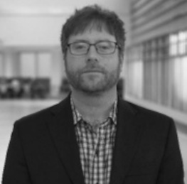 Over the past decade, however, researchers and students from Dalhousie’s School of Biomedical Engineering have been pushing the boundaries in brain cancer treatment. Led by Dr. Jeremy Brown (seen left), the team has designed the world’s first high-resolution endoscopic surgical and imaging probe. It uses an ultrafast imaging platform to allow surgeons to see brain tumours with ten times the resolution of conventional imaging such as MRI and CT scans. The 3mm-by-3mm device is inserted through a small keyhole created in the skull and allows surgeons to follow an exact path to the tumour so that it can be surgically removed.
While the probe itself is already a first of its kind, Dr. Brown is now working on a secondary feature for the device — a therapeutic tool that will non-invasively vaporize cancerous tissues with high-intensity sound waves.
“You’ll be able to image and treat tumours at the same time. This is ground-breaking research,” he says. “These brain tumours are universally fatal, so the question now is: can we cure an incurable cancer?”
Dr. Brown, who is an associate professor in the School of Biomedical Engineering and the Department of Electrical and Computer Engineering, was inspired to create the device after his PhD supervisor was diagnosed with a brain tumour over ten years ago.
“My PhD. Supervisor, Dr. Geoff Lockwood, was diagnosed with a glioblastoma brain tumour and given just 1-2 years to live following surgery,” says Dr. Brown. “When he told me that his cancer was terminal, I couldn’t believe that there was nothing that could be done. As a result, I started to look into this particular pathology more and more to see what improvements in treatment and patient survival could be made.”
More precision in treatment
Today, Dr. Brown and his group of graduate and undergraduate students have begun conducting tests with the therapeutic probe. The device uses sound to induce bubbles in tumors that mechanically rip cancer cells into small fragments. It also offers a much greater level of precision and effectiveness than other forms of treatment such as radiation therapy.
Dr, Brown is also collaborating with immunotherapy professors at Dal to look at how the body’s immune system could respond to this new form of ultrasound ablation.
“When tumours are disrupted, they release antigens that trigger an immune response, so the next step it to ablate some of these tumours and then combine it with immunotherapy,” he says. “There is have been several preclinical studies and one clinical study for liver cancer by groups outside of Dal that have demonstrated the positive immune response from this new therapeutic technology. After ablating primary liver tumours, all of the other secondary liver tumours shrank because of the stimulated the immune system.”
Over the past year, students enrolled in the School’s Certificate of Biomedical Engineering program have been working on a variety of applications to improve the efficacy of the device. This includes developing new micro-fabrication processes to build ultrasonic imaging and therapy devices.
“Undergraduate student groups have worked on developing new electronic hardware for the imaging and therapy system and the odd group would work on developing software for the imaging probes,” says Dr. Brown. “I have had really stellar students from Electrical and Computer Engineering and most of my graduate students are recruited from this program.”
Dr. Brown says students working on the research project benefit from the school’s unique, multidisciplinary training environment, which he adds is viewed as an enormous asset by industrial and academic recruiters.
“All these skills are highly sought after in growing sectors of the economy such as the semiconductor and med-tech industries,” he says. “Most previous graduates from my lab have gone on to work in fast-growing start-ups developing new biomedical devices.”