News
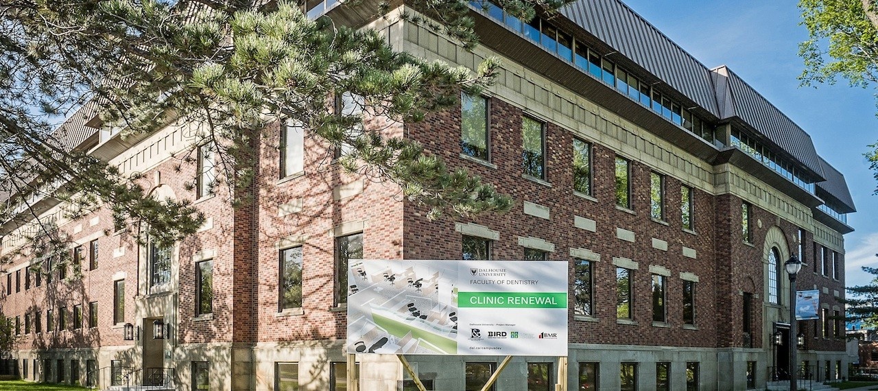
» Go to news mainThe brave new world of pre‑clinical labs
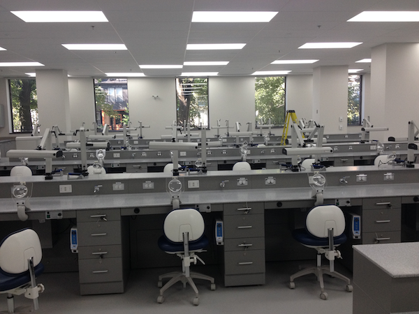 The new bench lab opened in November 2015
Venture into one of the new labs on Level 3 of the Dentistry Building and you would be forgiven for thinking that you have somehow stumbled into the future. The labs are sleek and uncluttered, beautifully lit, and sublimely clinical in white and grey.
The bench lab was completed in November 2015 and the simulation lab was completed in March 2016. Even though not all instructors have had a chance to teach in them yet, those who have speak highly of their features and the benefits they offer students.
Dental hygiene professor Peggy Maillet says: “What is really exciting about the labs is that you can lecture and have students apply the skill immediately.”
Bench lab
Each lab bench has:
- a task light
- knee-controlled electric lab motor with variable speeds
- a lab hand-piece
- an individually controlled vacuum unit with removable screened suction hood and magnifier
- a propane spigot
- a compressed air syringe
- two ethernet ports
The bench lab also has an AV podium, which controls numerous flat-screen monitors that the students can watch during demonstrations. The Prosthodontic Olympics, the annual student competition in Dr. Robert Loney’s Removable Prosthodontics II course, was held in the new bench lab and it proved a popular venue.
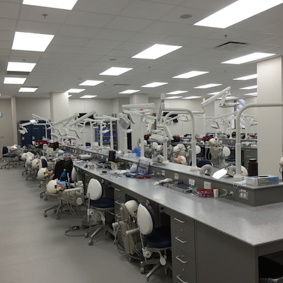
Simulation lab
The new simulation lab replaces the main treatment clinic for pre-clinical activity and is equipped with 50 A-dec simulators. Prior to the new lab, students worked on typodonts (mannequin heads) in the clinic, whereas the new simulators comprise a head and a torso, which gives them a better simulation of what it is like to work on a real patient. Each simulator also has a touchpad for adjusting and controlling the hand-pieces and operating the state-of-the-art dental light.
The simulation lab has many flat-screen monitors for viewing demonstrations. Two Planmeca radiography units use a direct digital sensor to provide instant digital intra-oral x-ray images of preclinical endodontic radiographs on each student’s laptop – quickly and safely, within the classroom. A third Planmeca radiography unit is planned for 2017.
A preclinical dispensary, located in between the two labs, supports both labs.
“With all the DDS2 students located in the same simulation lab space,” says Dr. Wayne Garland, who teaches Cariology II, Endodontics II, QPSM Cariology B and QPSM Endo in the lab, “it is much easier to provide preclinical instruction, as well as observe and provide support to all the students, as all the instructors are in one space.”
Dr. Cynthia Andrews, who teaches periodontology in the lab, agrees: “It allows for better calibration of instructors and more consistent messaging to students, since we are all in the same space and not spread out like we were in the clinic. The equipment is the same for all students and adjustable for all body types and sizes. They can develop ideal ergonomics and operating positions, which they can take with them into their clinical and patient care years. Finally, we can perform the majority of perio preclinical procedures in the sim lab, including ultrasonic instrumentation – a first, thanks to the simulator mannequins.”
Recent News
- Kendra Cleary: Recipient of the 2026 Dr. D. S. Precious University Medal in Dentistry
- Two grads, one mission: Serving as dentists in the Canadian Armed Forces
- "I can see myself doing this": Dalhousie Dentistry at PLANS Health Professions Exploration Day
- New Dentistry Building entrance lobby is unveiled and celebrated
- Vanessa Dairo‑Singerr: A focus on medically compromised patients
- Nadine Ayoub receives Impact Award
- Proud to support the Black Health Researchers' Hub
- Prof Shauna Hachey receives Golisano Health Leader Award 2026